


What is the story about?
What's Happening?
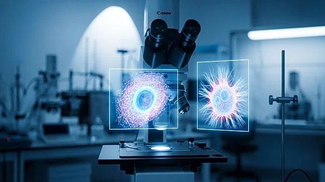
A recent study published in Light: Science & Applications has demonstrated the use of holo-tomographic flow cytometry (HTFC) for label-free phenotyping of acute myeloid leukemia (AML) blasts. The research, led by Pirone and colleagues, extends HTFC techniques to link genotype with observable cellular phenotypes without the need for labeling. This method utilizes holograms of rotating cells to reconstruct their three-dimensional refractive index maps, allowing for the detection of subtle nuclear shape differences associated with NPM1 mutations. The study introduces a new segmentation approach, concave-CSSI, which enables quantitative analysis of nuclei with cup-like morphologies. Additionally, virtual reality environments are integrated to enhance the interpretability of 3D data, offering immersive exploration of reconstructed tomograms.
Why It's Important?
This advancement in label-free holographic cytometry is significant for the medical field, particularly in the diagnosis and treatment of acute myeloid leukemia. By providing a non-invasive method to capture intrinsic structural properties of live cells, the technique could improve diagnostic accuracy and patient outcomes. The ability to detect statistically significant shifts in cellular architecture without staining offers a promising alternative to traditional fluorescence microscopy, which relies on exogenous dyes. The integration of virtual reality for data visualization may also support educational and clinical applications, potentially transforming how medical professionals interact with complex biological data.
What's Next?
Future developments may focus on enhancing the precision of single-cell classification, especially for diagnostically ambiguous samples. The study suggests integrating HTFC with complementary methods like correlative fluorescence imaging to reinforce its utility. Additionally, the introduction of standardized 'rolling-phantoms' could help consolidate resolution claims and promote adoption across laboratories. The potential use of artificial intelligence and machine learning for organelle-level segmentation is also highlighted, indicating a direction for further research and application in clinical settings.
Beyond the Headlines
The study's approach to label-free cytometry could have broader implications for the field of quantitative phase imaging, potentially influencing the development of new diagnostic tools and techniques. The use of virtual reality for data exploration represents a shift towards more interactive and intuitive methods of engaging with high-dimensional biological data, which could enhance both research and educational experiences.