What's Happening?
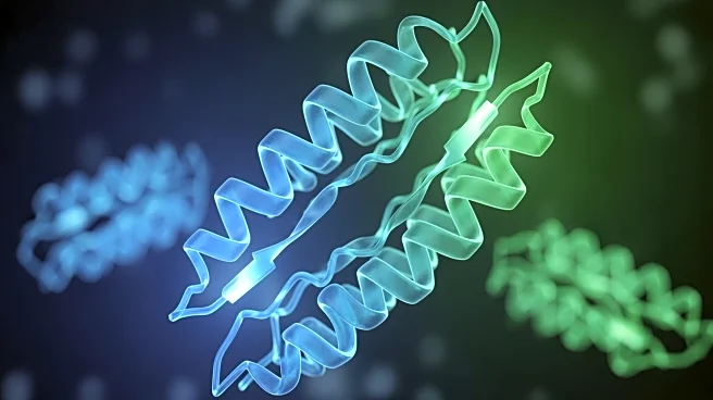
Researchers from Johns Hopkins University and Ludwig-Maximilians-University Munich have published the first 3D details of the ZAK protein's structure, a key component in cellular stress response. ZAK proteins regulate stress responses to various agents,
including UV irradiation, by interacting with ribosomes during protein translation. The study reveals molecular details of the ZAK protein, which could lead to the development of specialized treatments targeting this signaling network. The research involved engineering human cells to overproduce inactivated ZAK proteins, inducing ribosome collisions to activate ZAK. Using cryo-electron microscopy, scientists visualized the protein's structure, uncovering interactions with ribosomes that activate ZAK during collisions. This discovery provides insights into kinase proteins, which act as on-off switches in cellular processes, and could lead to more specific drug development with fewer side effects.
Why It's Important?
The detailed understanding of the ZAK protein's structure is a significant advancement in cellular biology, potentially leading to the development of targeted therapies for diseases related to cellular stress responses. By identifying specific interactions between ZAK and ribosomes, researchers can design drugs that precisely target these sites, reducing side effects associated with kinase inhibitors. This research could impact the pharmaceutical industry by providing a framework for developing new treatments for conditions involving cellular stress, such as cancer and neurodegenerative diseases.
What's Next?
The research team plans to capture more of ZAK's structure to further understand its activation mechanisms and functions when not connected to ribosomes. Future studies may explore the role of ZAK in other cellular processes, potentially leading to broader applications in drug development. Collaborations with pharmaceutical companies could expedite the creation of new therapies targeting ZAK and similar proteins.


