


What's Happening?
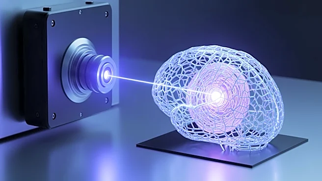
Researchers at MIT have discovered a novel effect in optical physics that could revolutionize the way living tissue is imaged. By manipulating laser light under specific conditions, they have managed to create a highly focused 'pencil beam' that can produce
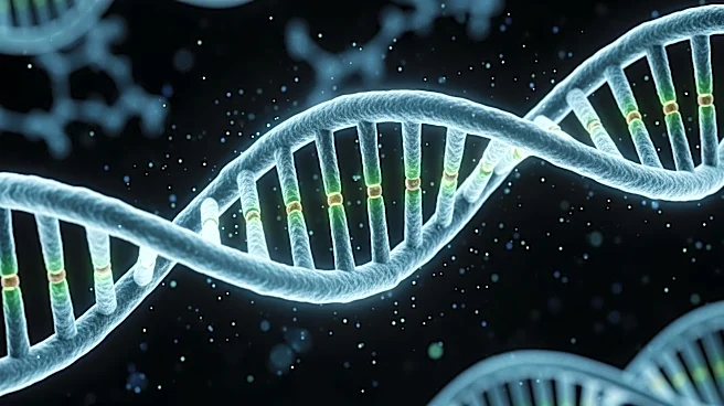
3D images of the human blood-brain barrier at speeds 25 times faster than current methods. This technique allows for real-time observation of drug absorption by individual cells, potentially improving the evaluation of treatments for neurological conditions like Alzheimer's and ALS. The research, led by Sixian You and published in Nature Methods, highlights a significant advancement in bioimaging technology.
Why It's Important?
This development is significant for the medical and pharmaceutical industries, as it offers a more efficient method for studying the blood-brain barrier, a critical factor in drug delivery for brain diseases. The ability to observe drug interactions in real-time could lead to more effective treatments and a better understanding of how drugs penetrate brain tissue. This could accelerate the development of therapies for conditions that currently have limited treatment options. Additionally, the technique's ability to produce high-resolution images quickly could enhance research capabilities in various biological and medical fields.
What's Next?
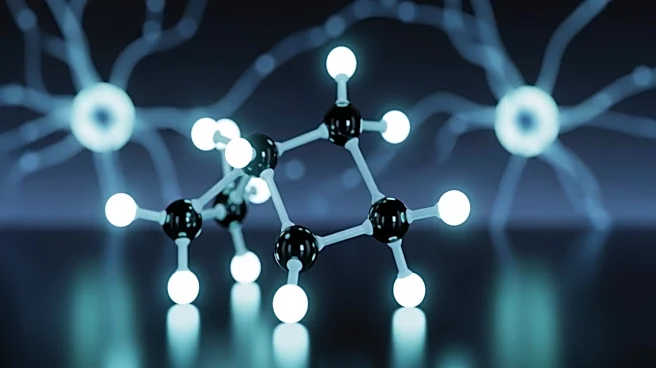
The MIT team plans to further explore the physics behind the self-organizing laser beam and extend the method to other applications, such as imaging neurons. They aim to refine the technology for practical use, potentially broadening its application in medical diagnostics and research. The research is supported by various institutions, including the National Science Foundation and MIT startup funds, indicating a strong foundation for future advancements.