
What's Happening?
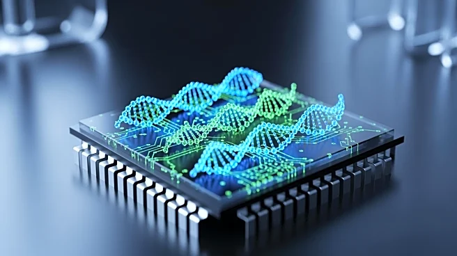
Researchers at Utrecht University have developed a fluorescent sensor that allows scientists to observe DNA damage and repair in real-time within living cells. This breakthrough, detailed in Nature Communications,
enables experiments that were previously impossible, offering a continuous view of DNA repair processes. The sensor, built from natural protein components, highlights damaged DNA without interfering with cellular repair mechanisms. It has been successfully tested in living organisms, such as the worm C. elegans, demonstrating its applicability beyond laboratory settings. The sensor can map DNA damage locations and assess protein interactions, potentially revolutionizing cancer biology, drug safety studies, and aging research.
Why It's Important?
The development of this sensor is significant for medical research, particularly in fields like cancer therapy, where understanding DNA damage and repair is crucial. Traditional methods required killing cells to observe repair events, but this sensor provides a continuous view, enhancing data accuracy and resolution. It could improve the precision of drug development and cancer treatments by offering real-time insights into DNA damage. The sensor's ability to work in living organisms opens new avenues for studying natural aging and exposure to mutagenic factors, potentially leading to advancements in clinical research and therapies.
What's Next?
The sensor's availability online allows immediate use by researchers worldwide, potentially accelerating discoveries in DNA repair mechanisms. As interest grows, further applications in clinical settings and drug development are expected. Researchers may explore its use in mapping DNA damage across genomes and testing how DNA location influences repair. The sensor's integration into existing research frameworks could lead to more efficient and cost-effective studies, enhancing our understanding of genetic diseases and treatment responses.