
What's Happening?
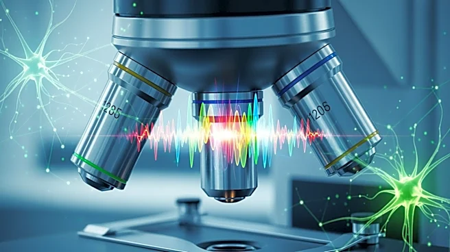
A new study published in Nature Communications Medicine reveals a diagnostic strategy combining hyperspectral imaging (HSI) with artificial intelligence (AI) to detect oxidative stress in red blood cells.
This non-invasive method identifies biochemical changes that alter membrane optical scattering properties, enabling early disease detection. The study demonstrates the method's ability to distinguish children with Autism Spectrum Disorder (ASD) from neurotypical controls, highlighting its potential for personalized diagnostics. The integration of lipidomic analysis and AI models provides a comprehensive framework for assessing oxidative stress and related cellular changes.
Why It's Important?
The ability to detect oxidative stress non-invasively represents a significant advancement in diagnostic technology. Oxidative stress is implicated in various diseases, and early detection can lead to more effective interventions. The study's findings suggest that hyperspectral imaging could be used for early diagnosis of neurodevelopmental disorders like ASD, as well as other conditions linked to oxidative stress. The integration of AI enhances the accuracy and efficiency of the diagnostic process, offering a promising tool for personalized medicine and continuous monitoring of cellular health.
What's Next?
Future research may focus on validating the hyperspectral imaging approach with larger and more diverse populations. The method's application could extend to a wide range of diseases associated with oxidative stress, including cardiovascular and metabolic disorders. The integration of optical imaging, lipidomics, and AI creates a platform for personalized medicine, supporting targeted interventions to restore cellular health. As biophotonic technologies advance, this study opens opportunities for translating innovative diagnostic methods into routine clinical practice.